

The physiology and treatment of frozen shoulder
What this diagnosis reveals about inflammation, fibrosis, and systemic health

I've been sidelined from surfing these past few months by shoulder pain. When I finally had it evaluated, the diagnosis was frozen shoulder, a common condition in women during midlife and perimenopause. While I have great respect for the physician, my right eyebrow raised at how quickly the explanation ended there. Perimenopause may be part of the story, but I didn’t want to believe it was the whole story. As it turns out, attributing frozen shoulder to perimenopause isn't exactly wrong, but it is incomplete in a way that is telling to my overall health.
Frozen shoulder is the common diagnosis, but it is clinically known as adhesive capsulitis. We will just call it AC for brevity hereon out. A much less comical-sounding diagnosis if you ask me. It affects a very small percentage of the general population, just 2- 5%, but is more prevalent (and rising) in patients with diabetes, thyroid disease, or autoimmune conditions.
It’s also frequently misattributed because rotator cuff tears, calcific tendonitis, and glenohumeral osteoarthritis can all mimic early presentations. The true diagnostic marker of restricted active and passive external rotation is easy to skip past in a busy visit.
The case I want to make is that estrogen decline is a real contributor, but treating it as a whole explanation causes clinicians to stop seeking further. And this is exactly the point where a fuller workup could catch something else of importance.
Anatomy and Physiology of AC
This is not merely an issue of a tight shoulder that needs to be stretched. It is pathologic tissue remodeling classically divided into three overlapping stages of freezing, frozen, and thawing.
Freezing. Pain is usually the dominant feature. Nighttime pain is common, movement becomes increasingly uncomfortable, and range of motion progressively declines. Lasts ~2 - 9 months.
Frozen. Pain may become less intense, but stiffness is more prominent. Activities such as dressing, reaching behind the back, fastening a bra, putting on a jacket, washing hair, or reaching can become remarkably difficult. Lasts ~4 - 12 months.
Thawing. Pain continues to recede, and motion gradually improves, sometimes over many additional months.
Patients do not always progress linearly through each stage of freezing, frozen, thawing. Some shoulders improve relatively fast, while others remain symptomatic for two or three years. Some recover near-normal motion, while others retain measurable limitations. Diabetes and secondary AC following surgery, trauma, or prolonged immobilization are generally associated with a more resistant course.
While the shoulder may be where the condition becomes visible, the course of AC is actually shaped by the interaction of local tissue biology, systemic physiology, pain processing, and the treatment environment surrounding the patient.
Let’s talk about what actually freezes.
The shoulder is a ball-and-socket joint, but the socket is quite shallow. It has an extraordinary range of motion that depends on a thin connective-tissue capsule that surrounds the joint while allowing the humeral head to move freely. Several areas are particularly important here:
Rotator interval - located between supraspinatus and subscapularis tendons
Coracohumeral ligament - regulates external rotation
Axillary recess - a fold in the lower capsule that unfolds as the arm elevates
In AC, these tissues become inflamed, thickened, fibrotic, and contracted. The capsule no longer expands normally, and joint volume decreases. External rotation is commonly the first and most severely restricted movement because the rotator interval and coracohumeral ligament are often prominently involved.
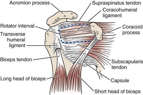
The restriction is therefore not merely a consequence of weak muscles or fear of movement. Eventually, there is a genuine mechanical barrier: the container surrounding the joint has become smaller and less compliant. This explains the clinical hallmark of restriction being both active and passive range of motion.
A patient cannot lift or rotate the arm normally, and the examiner cannot move it normally, either. In many rotator cuff disorders, active movement is impaired because movement hurts or the tendons cannot generate sufficient force, but passive range remains comparatively preserved. In frozen shoulder, passive external rotation is characteristically restricted as well.
The biological sequence is that inflammation then becomes fibrosis.
This diagnosis is best understood as an inflammatory-fibrotic continuum. In the earlier phases, the synovium and capsule become inflamed. Synovial hyperplasia, increased vascularity, immune-cell activity, and inflammatory signaling contribute to pain and irritability of the joint. A patient may still have a meaningful amount of motion, but movement (especially rapid or end-range movement) can be intensely painful.
Inflammatory mediators identified in frozen-shoulder tissue include:
IL-1β (interleukin-1 beta)
IL-6 (interleukin-6)
TNF-α (tumor necrosis factor alpha)
TGF-β (transforming growth factor beta)
The precise initiating event is often unknown, but the tissue environment increasingly favors fibroblast activation and extracellular-matrix production. Their normal function is to produce and maintain connective tissue; then during repair, they are tasked with synthesizing collagen and other components of the extracellular matrix. In frozen shoulder, fibroblast activity becomes excessive and poorly resolved.
Some fibroblasts transition into myofibroblasts, which pull on and contract the surrounding tissue, which helps explain why the capsule becomes both thicker and physically shorter. TGF-β is particularly relevant because it promotes fibroblast-to-myofibroblast transition, collagen synthesis, and matrix accumulation. At the same time, the balance between matrix production and matrix breakdown becomes disturbed. The result is excessive collagen deposition, extracellular-matrix stiffening, and capsular contracture.
This inflammatory-fibrotic continuum explains the clinical pattern that’s easy to miss: aggressive stretching during a freezing phase tends to provoke more pain and, plausibly, more fibrotic response.
Pain can be disproportionate to imaging results
Pain can persist even as motion improved in the thawing phase. Recent quantitative sensory testing work shows this isn’t purely local. Patients with AC demonstrate widespread hyperalgesia and altered pain processing that extend well beyond the shoulder itself. This is alongside autonomic symptom differences and central sensitization inventory scores that outpace controls.
This tracks with earlier findings that pain catastrophizing, hypervigilance, and spatial spread of pain correlate with sensitization measures in this population. Because the nervous system itself becomes more sensitive all over the body, the pain can linger even after the full range of motion returns.
A chronic, poorly resolved inflammatory process is a well-documented driver of central sensitization. Treatments aimed only at the joint (e.g., an injection, a series of stretches) can underperform if sensitization has taken hold and isn’t addressed alongside the mechanical issue. This is also a reasonable place to screen for depression and anxiety, both of which are prognostic for disability and pain severity in this population. It’s also a practical argument for pairing manual and exercise therapy with brief pain-neuroscience education, particularly in patients with a long or atypical course, and treating only the capsule in a sensitized system is treating half of the problem.
That same chronic, low-grade inflammatory load doesn't stay contained to the shoulder capsule either because thyroid, glycemic, and metabolic dysfunction all sustain their own versions of it elsewhere in the body. This raises the question of whether a sensitized nervous system and an unaddressed metabolic picture are reinforcing each other.
A systemic immuno-metabolic picture beyond “it’s menopause”
AC disproportionately affects women aged 40-60, and estrogen decline is a genuine part of the mechanism because falling estrogen weakens anti-inflammatory, antifibrotic, and antioxidant defenses that otherwise restrain capsular fibrosis. But a recent narrative review in the Journal of Clinical Medicine (2025) makes a case worth taking seriously: menopause is one input into a broader immuno-endocrine-metabolic disorder, not the whole explanation. Not every postmenopausal woman develops AC, and it also occurs in men, so deficiency alone does not account for the pattern.
The 2025 review also discusses the other axes converging on the same fibrotic endpoint:
Thyroid dysfunction, particularly hypothyroidism, independently promotes fibrosis via TGF-β activity, impaired mitochondrial function, and reduced matrix metalloproteinase expression. Even subclinical hypothyroidism has been implicated, which is easy to miss on a standard panel.
Hyperglycemia and insulin resistance. Elevated HbA1c is a consistent feature in AC cohorts. Chronic hyperglycemia drives advanced glycation end-product (AGE) accumulation, which cross-links collagen, stiffens the extracellular matrix, and activates RAGE-NF-κB inflammatory signaling, which then compounds the same fibrotic cascade described above.
Dyslipidemia. Elevated LDL and total cholesterol show up repeatedly in AC cohorts, both as a marker and a plausible mechanistic contributor (cholesterol is also the precursor for estrogen synthesis, so dyslipidemia and estrogen insufficiency can reinforce each other).
Endothelial dysfunction. Poor diet, AGEs, and oxidative stress impair nitric oxide signaling and vascular integrity, contributing to local microvascular inflammation in the capsule itself.
Circadian disruption and sleep. Estrogen, growth hormone, cortisol, and melatonin all follow circadian rhythms tied to tissue repair and inflammatory resolution. Fragmented sleep and light-at-night exposure impair this coordination and are an underrecognized contributor to the chronic low-grade inflammation sustaining AC.
Metabolic syndrome and adipose dysfunction more broadly, as visceral adiposity drives pro-inflammatory adipokine secretion (leptin and resistin) and reduced adiponectin, further impairing estrogenic receptor signaling independent of circulating hormone levels.
The throughline here is that almost all of these converge on the same downstream biology regardless of which upstream driver started the cascade: TGF-β activation, NF-κB signaling, impaired estrogen receptor function. So when a diagnosis of AC is provided, it’s worth inquiring about what else might be part of the picture.
What the research shows
Menopause doesn’t necessarily rule out a comorbid metabolic issue. The literature is fairly clear that metabolic syndrome prevalence rises through the menopausal transition. The 2008 SWAN study (Study of Women’s Health Across the Nation), a 9-year longitudinal cohort, found the increase tracks specifically with menopause versus just chronological age.
For any AC patient, published work supports a baseline look at:
TSH and free T4 (not TSH alone) - subclinical hypothyroidism has specifically been implicated, and would be missed by TSH screening thresholds alone
Fasting glucose or HbA1c - elevated HbA1c is a consistent feature across AC cohorts, and diabetic patients are well-documented to have a more resistant, longer course
Lipid panel - dyslipidemia recurs across the literature both as a marker and, plausibly, a mechanistic contributor
The case for a more extended workup gets stronger with specific features: bilateral presentation, onset in men, onset under 40, or a course not responding to standard conservative management past 4-6 weeks. In that subset, the literature also supports:
HOMA-IR alongside glucose and HbA1c
Metabolic markers including waist circumference, blood pressure, fasting triglycerides, hs-CRP, total cholesterol to HDL ratio
Sleep and circadian patterns - less a lab value than a history worth taking, given the role of circadian misalignment in sustaining low-grade inflammation
None of this is a substitute for standard orthopedic management, of course, but a reason the diagnosis has been treated in the literature as a potential window into broader metabolic health.
This workup runs alongside treating the shoulder, not instead of it. The shoulder itself still follows its staged course, and the right intervention changes considerably depending on which stage a patient is in:
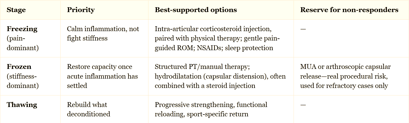
The trial evidence behind each treatment is worth a closer look, so let’s dive in.
Trial evidence: what it shows and where it’s thin
Corticosteroid injection has the deepest evidence base and remains a reasonable default, particularly early. A 2026 randomized trial found corticosteroid outperformed platelet-rich plasma (PRP) on pain and disability, with benefit holding through six months. Injection isn’t uniformly superior to physical therapy, however. A multimodal physiotherapy comparison against corticosteroid injection found physical therapy actually edged out injection on function and internal rotation.
Combining approaches tends to beat either one alone. One trial found hydrodilation plus corticosteroid plus physical therapy outperformed physical therapy by itself on nearly every measure. The summary here is less about which single treatment wins, but more about how injection and physical therapy are complementary, not competing.
Evidence quality on adjunctive and multifactorial approaches varies quite a bit. A systematic review and meta-analysis of multifactorial rehabilitation (combining manual therapy, exercise, and patient education) found benefit over single-modality approaches. Acupuncture-related therapies show comparative effectiveness in network meta-analysis, though heterogeneity across studies is substantial. Photobiomodulation (650nm laser) has plausible anti-inflammatory and pro-repair mechanisms, but the evidence base is still developing.
The practical takeaway
Frozen shoulder is a useful case study precisely because it resists the single-cause framing patients (and sometimes clinicians) default to. The capsule is where the process becomes visible, but the biology driving it (inflammation, fibrosis, pain sensitization) is shaped by thyroid status, glycemic control, lipid profile, circadian health, and hormonal status together, not any one of them alone. Treatment works best when it respects the stage a patient is actually in, injection and rehab together rather than as competitors.
And "it's hormonal" is rarely the whole story, whether or not the presentation looks textbook.
Alright then, I’m off to get my labs done and stare at the ocean for some mind-surfing.
References
Achilova, F., Daher, M., Nassar, J. E., Daniels, A. H., & Abboud, J. A. (2026). Frozen shoulder: Diagnosis and treatment of adhesive capsulitis. The American Journal of Medicine, 139(5), 598–605.
Hamed-Hamed, D., Pérez Montilla, J. J., Brindisino, F., Struyf, F., & Navarro-Ledesma, S. (2026). Integrative approaches to the understanding and management of frozen shoulder: A systematic review and meta-analysis of multifactorial interventions. Pain Research and Management, 2026, Article 3820129.
Hassan, M., Al Balah, O., & Osama, M. (2025). Physiological action of photobiomodulation using 650 nm diode laser for treating frozen shoulder. Lasers in Medical Science, 40, Article 520.
Janssen, I., Powell, L. H., Crawford, S., Lasley, B., & Sutton-Tyrrell, K. (2008). Menopause and the metabolic syndrome: The Study of Women's Health Across the Nation. Archives of Internal Medicine, 168(14), 1568–1575. https://doi.org/10.1001/archinte.168.14.1568
Ji, R., Huang, W., Weng, M., & Zhang, M. (2025). Comparative effectiveness of acupuncture-related therapies for frozen shoulder: A systematic review and network meta-analysis. Frontiers in Medicine, 12, Article 1673193.
Li, D., St Angelo, J. M., & Taqi, M. (2025). Adhesive capsulitis (frozen shoulder). In StatPearls. StatPearls Publishing.
Navarro-Ledesma, S. (2025). Frozen shoulder as a systemic immunometabolic disorder: The roles of estrogen, thyroid dysfunction, endothelial health, lifestyle, and clinical implications. Journal of Clinical Medicine, 14(20), Article 7315.
Wang, S., Zhang, F., Chen, J., & Fu, G. (2025). Tuina combined with other treatment methods for scapulohumeral periarthritis (frozen shoulder): Bayesian network meta-analysis. Journal of Pain Research, 18, 4673–4686.
Yu, S., Gou, L., Huang, J., Wang, L., Chen, B., Kang, Y., & Yang, L. (2026). Frozen shoulder is more than local pain: Widespread sensory hyperalgesia or hypoesthesia? European Journal of Pain, 30, Article e70215.
Zare, M. A., Ebrahimzadeh, M. H., Moradi, A., Zeinalzadeh, A., & Nazary-Moghadam, S. (2026). Clinical outcomes of intra-articular corticosteroid injection vs. multimodal physiotherapy in patients with frozen shoulder in short term: A randomized clinical trial. Scientific Reports, 16, Article 3607.